Ursaki Nataliia
16 years
chief physician, Ph.D. in Medicine, ultrasound doctor, obstetrician-gynecologist
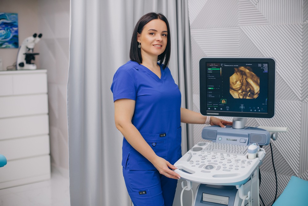
The modern and advanced ultrasound machine of the expert class, Voluson E8 Expert, allows obtaining the most accurate information about the embryo at all stages of development. The HD LIVE feature, four-dimensional ultrasound scanning using ultrasound waves, provides a clear, colored, and real-time image of the fetus starting from the 10th week of pregnancy. Compared to the regular black and white ultrasound in Ivano-Frankivsk, which is informative and understandable only for the doctor, this is a more precise picture and information due to the high clarity of visibility of all tissues and assessment of the condition of various body parts in three projections simultaneously.
This type of scanning, available in our city and region, enables us to see the fetus clearly: its smile, tongue, facial expressions, dancing and spinning movements, yawning, hand and foot movements, sucking fingers, or just sleeping. It opens up the possibility of counting the fingers on the hands and feet and conducting a thorough examination of the brain, spine, heart, and internal organs. Even with the incorrect placement of the fetus in the mother’s womb, by switching to another scanning method, we can assess the condition of the necessary organ. The light game, in which different shadows are overlaid, will show incredible images of your baby.
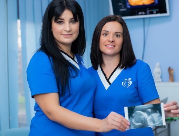
The examination will be understandable not only to the doctor but also to the future mother and father, whom we always invite to a full examination, which is important from a psychological and emotional point of view. Our doctors will answer your questions, determine the gender of the baby with 100% accuracy, and provide the opportunity to examine the fetus down to the smallest detail, using an additional imaging screen for the patient.
The 30-second video clips recorded during the examination can be transferred to a flash drive or digital photograph, which will preserve your first encounter in an album.
The 4D program is not just interesting to see the fetus and its movement in real-time, but its greatest advantage lies in the accuracy of detecting the development and possible abnormalities of the fetus. With a wide range of built-in programs, starting from the earliest stages of pregnancy (from 11-12 weeks), we qualitatively examine:

We give an accurate conclusion of the ultrasound examination with the indicated weight of the fetus (from 70 g) and calculate the predicted due date.
The Med-Atlant Medical Center in Ivano-Frankivsk follows the orders and protocols of the Ministry of Health of Ukraine, according to which pregnancy ultrasound screening is performed three times.
In addition to the three screenings mentioned above, your doctor may recommend additional testing – there is no need to be afraid of this, as ultrasound diagnostics is not harmful, thanks to the use of the latest technologies, and does not cause complications for both the mother and the baby!
| Service | Price (UAH) |
| Ultrasound diagnostics | |
| Ultrasound Examination “Preoperative Package” (abdominal organs, kidneys and bladder, mammary glands, thyroid gland) *designed for patients preparing for surgical intervention | 950 |
| Ultrasound Examination “Women’s Package” (transvaginal examination of pelvic organs with Doppler imaging, mammary glands, thyroid gland) | 1100 |
| Ultrasound Examination “Men’s Package” (abdominal organs, kidneys, prostate, urinary bladder) | 950 |

In an uncomplicated pregnancy, it is recommended to have 4 scheduled ultrasound scans.
Additional (unscheduled) ultrasounds may be advised by an obstetrician-gynecologist in case of complications or suspected pathologies. At our clinic, all ultrasound diagnostics are performed using new, expert-class equipment, which is completely safe for both the mother and the baby.
This means that pregnant women can undergo the number of ultrasound exams recommended by their doctor without risk.